In the ever-evolving world of ophthalmology, precision is paramount. Enter the DGH A Precision Ophthalmic Ultrasound Device—an innovation that promises to revolutionize how we diagnose and treat eye conditions. Whether you’re an experienced practitioner or just stepping into the field, this cutting-edge technology offers unparalleled accuracy and efficiency in ocular imaging. Say goodbye to guesswork and hello to crystal-clear insights! Join us as we dive into the remarkable features of the DGH A, exploring how it enhances patient care and transforms diagnostic capabilities for eye specialists everywhere. Get ready to see your practice—and your patients—in a whole new light!
Introduction to the DGH A and its various uses
The world of ophthalmology is constantly evolving, and precision tools play a pivotal role in enhancing patient care. Enter the DGH A, a state-of-the-art ophthalmic ultrasound device that has transformed how eye specialists diagnose and treat various ocular conditions. This innovative technology offers unmatched accuracy and versatility for visualizing the intricate structures of the eye.
From measuring intraocular tumors to assessing retinal detachment, the DGH A proves invaluable across multiple applications. As we dive deeper into its features, history, and real-life impact on patient outcomes, it becomes clear why this device is making waves in the field of eye care. Whether you’re an ophthalmologist looking to upgrade your equipment or simply curious about advancements in medical imaging technology, you won’t want to miss what makes the DGH A truly remarkable.
History and development of ophthalmic ultrasound devices
The journey of ophthalmic ultrasound devices began in the 1950s. Early pioneers sought to visualize internal eye structures using sound waves. This marked a significant shift in diagnostic methods.
By the 1970s, advancements led to A-scan technology, allowing for precise measurements of ocular components. It was a breakthrough for cataract surgery planning and intraocular lens calculations.
Throughout the years, B-scan ultrasound emerged as another vital tool. It provided cross-sectional images of the eye, enabling doctors to diagnose tumors and other anomalies effectively.
As technology progressed into the late 20th century, digital imaging transformed how practitioners approached diagnostics. Enhanced resolution improved accuracy and made examinations faster than ever before.
Today’s devices are more compact and user-friendly while offering advanced capabilities like color Doppler imaging. This evolution has significantly impacted patient care by aiding early detection and treatment strategies.
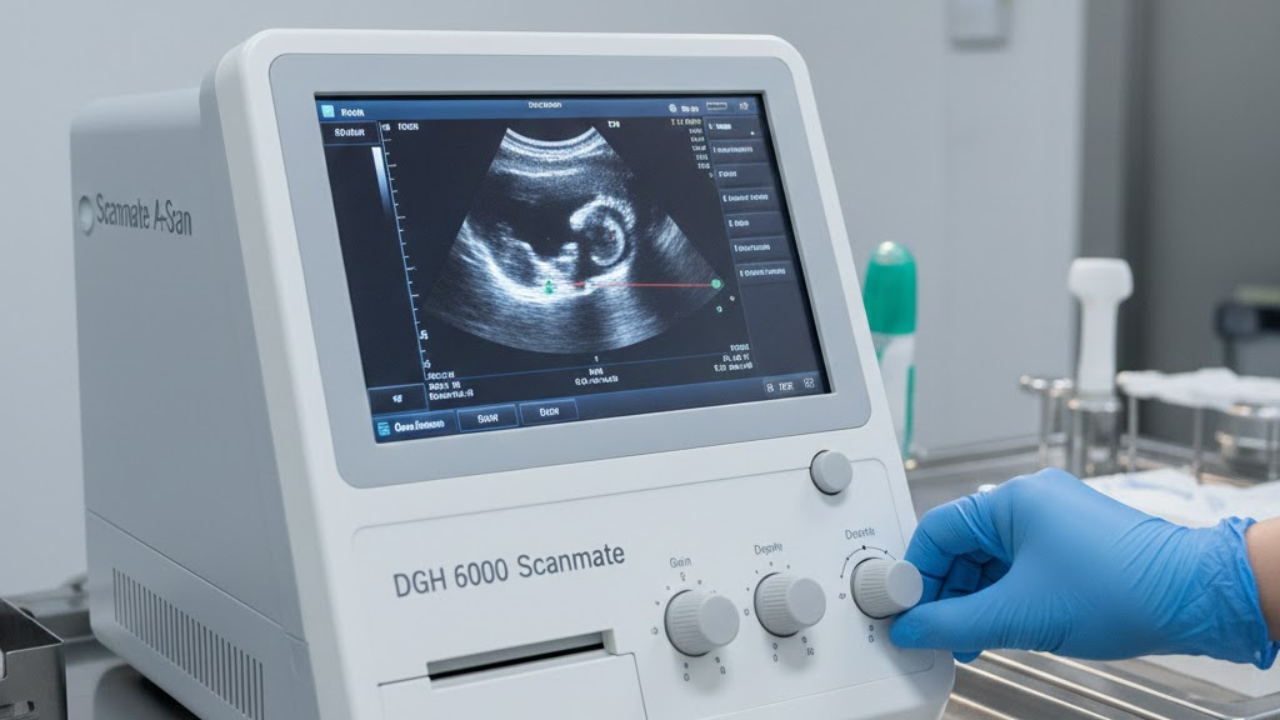
Features and capabilities of the DGH 6000 Scanmate A
The DGH 6000 Scanmate A sets a new standard in ophthalmic ultrasound technology. With its high-resolution imaging, it provides detailed insights into ocular structures. Clinicians can visualize the anterior and posterior segments of the eye with remarkable clarity.
This device features an intuitive interface, simplifying operation for medical professionals. Users can easily adjust settings to tailor examinations to specific patient needs. The lightweight design enhances portability, making it ideal for both clinic and hospital environments.
Equipped with advanced software, the Scanmate A facilitates automated measurements and calculations. This ensures accuracy while saving time during assessments. Additionally, real-time image processing allows practitioners to make immediate decisions based on their findings.
Compatibility with various probe types expands its versatility in clinical applications. From cataract evaluations to retinal assessments, the DGH 6000 is designed to adapt seamlessly across diverse ophthalmic practices.
Benefits and advantages of using the DGH A in ophthalmology
The DGH A revolutionizes the field of ophthalmology with its precision and efficiency. This device allows for accurate measurements, enhancing diagnostic capabilities significantly.
One major advantage is its user-friendly interface. Practitioners can quickly learn to operate it, streamlining workflows in busy clinics.
Another benefit lies in its portability. The compact design enables easy transport between different practice locations or within hospitals, ensuring that eye care can be delivered wherever needed.
Additionally, the DGH A provides high-resolution imaging. This clarity aids in identifying subtle changes in ocular structures, which is crucial for early disease detection and management.
Furthermore, the system’s versatility supports various applications—be it assessing glaucoma or evaluating retinal conditions—making it a valuable tool for any eye care professional seeking comprehensive diagnostic solutions.
Case studies and real-life examples of successful use of the DGH A
A notable case involved a patient with suspected retinal detachment. The ophthalmologist utilized the DGH A to obtain detailed images, confirming the diagnosis swiftly. This enabled timely intervention and saved the patient’s vision.
In another instance, a clinic used the DGH A for pre-operative assessments in cataract surgery. The precise measurements provided by this device helped surgeons plan their procedures more effectively, resulting in improved surgical outcomes.
Additionally, therapists working with pediatric patients have reported success using the DGH A to monitor conditions like congenital glaucoma. The compact design made it easier to engage young children during examinations.
These examples highlight how versatile and effective the DGH A can be across various scenarios in ophthalmology. Each case illustrates its ability to enhance diagnostic accuracy and patient care significantly.
Comparison with other ophthalmic ultrasound devices on the market
When comparing the DGH A to other ophthalmic ultrasound devices, a few key factors stand out. Many competing models lack the precision and clarity that the DGH A consistently delivers.
The user-friendly interface of the DGH A sets it apart from more complex machines on the market. This ease of use is essential for busy clinics aiming to maximize efficiency.
Another critical aspect is its portability. While some devices are bulky and difficult to transport, the DGH A offers convenience without sacrificing quality.
Moreover, technology integration varies widely among competitors. The DGH A features advanced software that enhances diagnostic capabilities through detailed imaging analysis.
Finally, customer support often makes a significant difference in operational success. Users frequently report positive experiences with DGH’s dedicated service team, which can be rare in this industry.
How to properly use and maintain the DGH A for optimal results
To maximize the performance of the DGH A, start with proper training. Familiarize yourself with its interface and functionalities through comprehensive user manuals and hands-on practice.
Regular calibration is essential for accuracy. Perform routine checks to ensure measurements are precise. This will help maintain consistency in your results.
Cleaning should not be overlooked. Use approved disinfectants on transducers and surfaces after each use to prevent contamination.
Store the device in a cool, dry place when not in use. Avoid exposure to extreme temperatures or humidity, which can affect its lifespan.
Promptly address any issues that arise during operation by consulting technical support or service professionals. Timely repairs can save you from larger problems down the line.
Lastly, keep track of software updates as they often include improvements and new features that enhance functionality.
Customer reviews and testimonials from users of the DGH A
Users of the DGH A often rave about its precision and reliability. Many ophthalmologists have noted how it enhances their diagnostic capabilities. The clarity of images produced is frequently highlighted as a key feature.
One user mentioned that the DGH A significantly reduced their examination time without compromising quality. They appreciated how easy it was to integrate into their existing workflow.
Another doctor shared a story about identifying subtle retinal changes using the device, which led to timely interventions for patients. Feedback like this underscores its impact on patient care.
Moreover, technicians value the intuitive interface. Training new staff becomes smoother, allowing them to focus more on patient interaction rather than struggling with complex equipment.
These testimonials paint an encouraging picture for any practice considering adopting this technology in their ophthalmic services.
Future advancements and potential applications for the DGH A technology
The future of DGH A technology is promising, with numerous advancements on the horizon. Researchers are exploring enhanced imaging capabilities that could provide even greater detail and accuracy in ocular assessments.
AI integration stands out as a game-changer. By leveraging machine learning algorithms, data analysis can become faster and more precise, allowing for earlier detection of conditions like glaucoma or retinal detachments.
Telemedicine applications are also being developed. Remote access to diagnostic results will enable eye care professionals to consult and make informed decisions without geographical limitations.
Moreover, there’s potential for miniaturization of the device. Compact models may soon allow for easier portability in various clinical settings, ensuring accessibility wherever needed.
Lastly, ongoing collaboration between engineers and ophthalmologists aims to refine user interfaces further. This partnership will help create tools tailored specifically to practitioners’ daily needs.
Conclusion: Why the DGH A is a game-changer in ophthalmic care
The DGH A stands out as a remarkable innovation in the field of ophthalmic care. Its precision and versatility make it an invaluable tool for eye specialists. With its advanced features, including high-resolution imaging and user-friendly interface, the device streamlines the diagnostic process.
Healthcare professionals are increasingly recognizing the benefits of incorporating this technology into their practices. Case studies illustrate how patients receive quicker diagnoses and more effective treatment plans thanks to timely insights provided by the DGH A.
As advancements continue within medical technology, tools like the DGH A pave the way for improved patient outcomes. The commitment to enhancing vision health is evident in every aspect of this device’s design.
For any ophthalmologist looking to elevate their practice, embracing innovations like the DGH A could truly transform patient care forever.